Article
Development of a Rapid and Convenient Method for Sampling Airborne Virus based on Nanoparticle Adsorption
Chun Meng*, Yulin Xiong, Weiliang Zhuang, Hang Wang, Xian’ai Shi, Yanghao Guo
Department of Bioengineering, College of Biological Science and Biotechnology, Fuzhou University, Fuzhou, Fujian 350108, China.
*Corresponding author. E-mail: mengchun@fzu.edu.cn, Tel: 86-591-22866379, Fax: 86-591-22866379
Citation: C. Meng et al. Development of a Rapid and Convenient Method for Sampling Airborne Virus based on Nanoparticle Adsorption. Nano Biomed Eng. 2010, 2(3), 171-176.
DOI: 10.5101/nbe.v2i3.p171-176.
Abstract
An improved aerobiological virus sampling method was developed based on adding adsorptive nanoparticles in samplers for concentrating viruses in sampling liquid buffers. The objectives of this research were to select effective adsorptive materials and optimize sampling parameters for increasing recovery of airborne viruses, such as influenza A virus or respiratory syndrome virus (RSV). Three kinds of polycation nanoparticles were evaluated for direct effects on absorption and desorption of influenza virus hemagglutinin and DNA. Chitosan particles showed good performance in absorption and desorption for both influenza virus hemagglutinin and DNA. A subsequent study evaluated the effects of collection buffer, pH and sampling time on the recovery of aerosolized viruses using a method for making direct comparisons of three treatments. The results demonstrated that various components in air-sampling collection buffer, impinger model, and sampling time, independently influenced the recovery of viruses. It was shown that adsorptive samplers with air disperser had the highest levels of sensitivity and repeatability in virus sampling. Both unspecifically adsorptive chitosan particles and specifically adsorptive particles labeled specific antibody to virus significantly enhanced recovery rate of aerosolized viruses. We succeeded to sample low level different pathogen viruses in outdoor environments with the optimized sampling system.
Keywords: Aerobiological virus sampling method; Adsorption; Polycation chitosan nanoparticles; Influenza virus; Air disperser
Since the beginning of last year, the H1N1 subtype of influenza A virus (2009 H1N1or SIV) has been circulating in America, and now in Asia, European coun- tries, causing widespread infection in Human beings. As of September 13 last year, the World Health Organ ization regions had reported more than 296,471 labora tory-confirmed cases of 2009 H1N1 influenza virus with at least 3,486 deaths (http://www.who.int/csr/don/2009_09_18/en/ ). With the season change to fall and winter, we will face the threat of H5N1 subtype of in- fluenza A virus in the northern hemisphere. In a very long period of time, the outbreak of influenza A virus will be a big threat to humans health, and millions of people worldwide will suffer from this disease and die from the pandemic Influenza. Infection by direct contact can occur when infected hosts are in close proximity with a susceptible popula- tion. Moreover, infected hosts can transmit the disease not through direct contact but through airborne transmission. Viruses, can remain infectious outside their hosts for prolonged periods of time, and this can lead to infections by indirect contact in confined or public space, such as public transport, meeting room, bar, and so on [1]. The probability of airborne transmission of an infectious disease can be determined by conducting epidemiological studies and/or by directly analyzing the virus content of air samples [2]. Though effective vaccines and drug treatments could in some degree make the prevention and control of this disease, more effective measures for limiting a potential outbreak is identifying and controlling the source of inuenza. One of the most effective prevention methods is a large-scale quarantine of suspected patients. But it is still not enough for controlling suspected patients because suspected patients meant that the man had been likely infected with the flu virus. If we detect the virus in the air effectively, we may manage to reach earlier prevention of virus transmission to susceptible people. Detecting virus in air is very essential because inuenza is a typical airborne transmission disease caused by influenza viruses. Impingers direct a converged stream of environmental air onto or in a liquid collection medium to recover airborne particles in the liquid phase of the collection system [3-10], Which can be used as virus sampling because it is more effective than filters, bubblers, or impactors for capturing airborne viruses [11-13]. But it is less effective in detecting low concentration viruses in air because of difficulty of concentrating viruses and inefficiency in trapping viruses in air. Development of a sensitive, quick, accurate, and comprehensive sampling system for air virus detection becomes very essential. In this study, we reported the development of a new liquid impinger sampling system for sampling influenza. A virus in air based on absorptive method in liquid.
Table 1. PCR primer and hydrolysis probe sequences
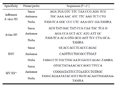
FAM, 6-carboxyfluorescein; TAMRA, 6-carboxytetramethylrhodamine.* World Health Organization. CDC protocol of real-time RT-PCR for swine influenza A (H1N1) http://www.who.int/csr/resources/publications/s wineflu/realtimeptpcr/en/index.html ).
2. Materials and Methods
2.1 Particles preparation
Chitosan nanoparticles were prepared according to the procedure reported by Calvo [14] based on the ionic gelation of CS with TPP anions. 201-4 anion exchange resin (strong basic quarternary ammonium) was purchased from Huachang Polymer Co., Ltd, China. DEAE Sephadex A-25 was purchased from Amersham Pharmacia. Here, an oxime-based chemistry method was used for covalently linking hemagglutinin antibody to polysaccharide particles. In this process, aldehyde carbonyls of polysaccharides are reacted with the highly nucleophilic aminooxy group to form oximes [13]. For the conjugate, the ratio (wt/wt) of polysaccharides/ antibody was at a range between 1:0.002 and 1:0.005.
2.2 Samplers and sampling methods
At a vacuum pressure of not more than -0.05 atm, the adsorptive sampler (a 0.3 liter glass cylinder reser- voir, 0.2 m high) operated at a flow rate of 1.5 liters per minute. And a circle polyethylene tube (inner diameter: 2.0mm, thickness: 0.5mm) containing full of holes on the wall of part immerged under water was used as the disperser. Vacuum pressure was maintained using oil- less pumps and was monitored using a vacuum pres- sure gauge. Flow rates of impingers in liters per minute were verified using a flow meter. A commercial impin- ger model was compared in terms of the recovery of aerosolized viruses. We used a similar equipment designed by Hermann to test the recovery efficiency of air virus [15]. A 20 liter glass reservoir was modified to allow simultane- ous sample collection at outlet ports. High pressure sterilization silicone tube was used to connect the sam- pling bottles to the outlet ports. This arrangement made it possible to test up to different treatments simulta- neously on the same cloud of aerosolized. Isolation of viruses from particles was performed with different concentration NaCl buffer (5 ml buffer per ml particles).
2.3 Nucleic acid isolation and amplification with RT-PCR and real-time PCR.
The influenza virus H1N1 (a gift from Prof. Pingfan Rao, Fuzhou University) DNA and membrane protein hemagglutinin was extracted with commercial kits purchased from Sangon, China, respectively. A QIAGEN QIAamp Viral RNA Mini kit was used for the extraction of nucleic acid from all of the samples. The starting material for nucleic acid isolation was 200µl viral isolate in dilution buffer (phosphate- buffered saline). RNA was eluted into 50µl water. The primer sequences used for these studies are shown as Tab 1. All probes were labeled at the 5’ end with the 6-carboxyfluorescein (FAM) reporter dye and at the 3’ end with the 6-carboxytetramethylrhodamine (TAMRA) quencher dye. The Qiagen one-step RT- PCR kit was used with a 20 l reaction mixture accord- ing to the reference [16]. The RT step conditions for all primer sets were 30 min at 50°C and 15 min at 94°C. PCR cycling protocol was used for the matrix gene primer set as described in table 1. Hemagglutinin was extracted from H1N1 viruseswith a membrane protein extraction Kit (Shanghai Sangon, China) according to the recommendations. The level of hemagglutinin was detected with ELISA men- tod [17]. Statistical analysis Data (including RSD value) were analyzed using the statistical software program (Excel).
Based on ion adsorption of viruses or cells with surface negative charges on polycations particles, we developed an adsorptive method through adding polycation particles as the adsorbent in impingers for sampling viruses in air. It was shown that all selected particles could effec- tively absorb both DNA and protein (data not shown) when DNA extract or membrane protein extract of in- fluenza virus was added in water. DEAE resin showed good performance in absorption and desorption of he- magglutinin (Fig.1), but it absorbed DNA too firmly to be eluted with 0.5M NaCl (Fig.2). Difficulty of eluting both DNA and protein from the resin 201-4 limited its application in the next study. It was shown that only chitosan particles of the three polycation absorption resins evaluated exhibited satisfying results on the both absorption and desorption of either virus DNA or hemagglutinin in the buffer. On the basis of the overall results, chitosan nanoparticles were selected for use in the following work.
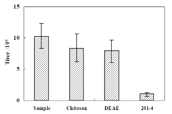
Figure 1. Collection of hemagglutinin by different particles in water.
Hemagglutinin was detected with ELISA. Eluate sample is eluted with 0.5 M NaCl. Both eluate and adsorbate were diluted to a same volume when detected the virus hemagglutinin level. The content per ml water of influenza virus hemagglutinin was extracted form 1000 copy/ml virus and the addition of chitosan particles was .0.05 g dry weight /ml.
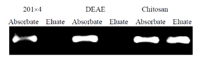
Figure 2. Collection of influenza virus DNA by different particles in water.
Virus DNA was detected with PCR. Eluate sample is eluted with 0.5 M NaCl. Both eluate and adsorbate were diluted to a same volume when detected the virus DNA level. The content per ml water of influenza virus DNA was extracted form 1000 copy/ml virus and the addition of chitosan particles was .0.05 g dry weight /ml.
Figure 3. Effect of components in buffer on virus adsorptive efficiency.
Virus was detected with real time PCR. Eluate sample is eluted with 0.5 M NaCl. Both eluate and adsorbate were diluted to a same volume when detected the virus level. The content per ml buffer of influenza virus was about 1000 copy/ml.
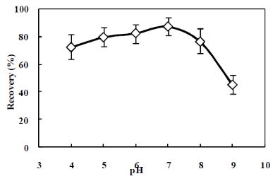
Figure 4. Effect of pH on virus adsorptive efficiency
Virus was detected with real time PCR. Eluate sample is eluted with 0.5 M NaCl. Both eluate and adsorbate were diluted to a same volume when detected the virus level. . The content per ml buffer of influenza virus was about 1000 copy/ml.
3.2 Effect of solutions and pH on virus adsorption efficiency in different buffer
The absorptive effect of the different content in the buffer on virus adsorption with chitosan particles was tested. Addition of 0.5% BSA significantly (P<0.01) reduced absorptive rate of viruses compared to water (Fig.3). Absorptive rate of 0.1 M PBS and 0.1 M NaCl buffer were not significantly different from the absorp- tive rate of water (P > 0.05). Chitosan particles showed good adsorptive result with 83% virus recovery when only water was used as adsorptive buffer. So water was selected for further evaluation in experiment of pH ef- fect on virus absorption. Fig.4 shows the virus absorption results of chitosan particles in the water at pH from 4.0 to 10.0 (adjusted with 0.1 M NaOH and 0.1 M HCl). There was no sig- nificant difference in virus absorption in the range from pH 5.0 to pH 8.0 with more than 80 % virus recovery. There was a significant decrease of virus absorptive efficiency when buffer pH exceeded the pH 5.0-8.0 range.
3.3 Study of sampling virus in air with adsorptive method
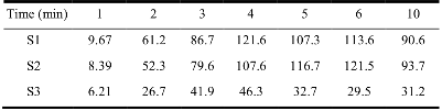
The average virus concentrations per release in the each trials was about 1.2´103 copy (the range was 0.9´103 ± 50 to 1.3´103 ± 50 copy/m3) per m3 of air, during release of viruses, the air rate through the room was 2.0 m3/min. In this test, an impinger instrument without adsorptive particles (S1) was used as the control. The results of experiments to sample viruses by the adsorptive method (including two samplers, one is impinger S2 and the other is impinger S3 with an air disperser) through addition of adsorptive particles and common impinger S1 for comparison were shown in Fig.5. As estimated by quantitative RT-PCR, the mean adsorptive saturation time (1000 copy virus/m3) was about 4 and 6 min and copies of virus collected across at saturation time point were 1.02×10³, 0.96×10³ copy/m3 for the S2 and S3 respectively. Recover rate of viruses reduced to 0.6×10³copy/m3 for S1 sampler without adsorptive particles at 10 min. Addition of adsorptive particles in the sampler collected a significantly greater amount of viruses and higher sampling peed than the common impinger sampler. Levels of repeatability were estimated for the three samplers according to relative standard deviation (RSD) value (Table 2). The results showed that the adsorptive sampler with disperser exhibited both the highest level of repeatability and the greatest level of sensitivity in the three experiments, followed by adoptive sampler without disperser. Nonhomogeneous distribution of gas resulted in some decrease in the level of repeatability in both the adoptive sampler without disperser and the common impinger sampler.
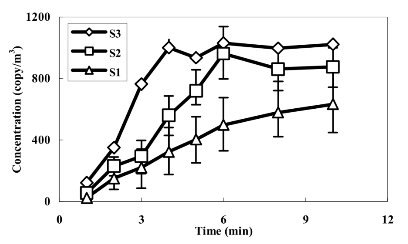
Figure 5. Collection of viruses in air with different samplers Each experiment was repeated five times
Table 2. The RSD value of each sample in Fig.5
3.4 Application of air sampling in different environments
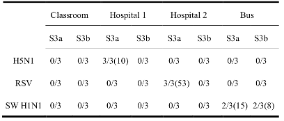
Aerobiological sampling conducted in hospitals, buses and classrooms in Fuzhou University, all located in Fuzhou city, was used to evaluate the usefulness of the adsorptive sampler with disperser in public environments. The trials were repeated three times in each sampling point and the sampling time was set to ten minutes. Low levels of airborne viruses were detected in our trials (Table 3). Based on estimated values by quantitative RT-PCR method, 10 copies H5N1, 15 cop- ies H1N1 and 53 copies respiratory syncytical virus (RSV) per m3was detected in a hospital near the Minjiang River, in a bus and in a hospital in downtown of Fuzhou respectively. We also tried to use particles labeled H1 antibody on particle surface through covalently linking proteins to polysaccharides as the absorbent to sample viruses in air. A similar sampling result (adsorptive efficiency) as chitosan particles was obtained. And the specificity of virus adsorption was improved after the nanoparticles were labeled with specific antibodies. S3a: chitosan particles was used as the adsorbent; S3b: particles labeled with H1 antibody was used as the adsorbent; the test was repeated three times at each site, and the numerator represented positive times. It was the average copies of viruses in brackets (copy/ m3).
Table 3. Virus detection results sampling from different public sites
In primary health care, the probability identification (or exclusion) of the pathogen causing respiratory symptoms should preferably be done in high risk area during the high season of flu. The development of sampling air virus method becomes very necessary be- cause many viruses infected the hosts through airborne transmission. Sampling airborne virus is still difficult and the part reason of difficulty of recovery of airborne viruses is that virus particles in air are too small to catch. Low concentration of airborne virus in the environment is another key restrictive factor. We can concentrate microorganism cells through membrane filtration method when concentrations of airborne microor- ganism cells in the environment are very low [18], but we hardly manage to concentrate viruses through filtra- tion method. So arresting viruses and concentrating viruses are two key factors for increase of sampling sensitivity. Polycation molecular could effectively adsorbed vi- rus particles which had negative surfaces through ion adsorption. The ability of the air-sampling system to recover airborne viruses could be improved by adding polycation particles. The results showed that the poly- cation resin exhibited strong affinity was not suitable to be used to sample viruses because virus components are hardly to elute from the resin (Fig.1). A resin with moderate affinity to viruses is a fit candidate as virus adsorbent. The results of the experiment showed that the addition chitosan nanoparticles can not only en- hance virus adsorptive efficiency but also easily release viruses when eluted with 0.1 M NaCl buffer (Fig.1, Fig.2). A suitable pH is very important for chitosan particles and viruses embedded negative charged bimo- lecular lipid membrane containing some specific mem- brane proteins to keep ion state. Chitosan are positively charged based on poly(ethyleneimine) and pH of ad- sorptive buffer is a key factor for virus adsorption. So- lutions with a pH less than 8.0 could make chitosan containing enough cation groups. pH 5.0-8.0 range is fit for both virus adsorption (keeping suitable charged groups on both sorbent and adsorbate) and virus elution from the sorbent. It had been demonstrated that addition of polycation chitosan nanoparticles could increase the sensitivity of the sampling virus method. According to the adsorptive results, the disperser could improve sampling sensitivity. The sampler with a disperser could make gas flow injected in liquid phase become small bubbles, which with high specific surface area could enhance contacted probability between virus in air and liquid or particles in liquid. The homogeneous bubbles provided a stable sampling environment which resulted in a high level of sampling repeatability. Labeled the specific H1 antibodies on particle surface could improve adsorptive specificity for influenza virus. Particles without labeling antibody were suitable to sample different viruses in air. In conclusion, the sampler can be used to sample airborne virues broadly or specifically. Both methods might be used based on different demands. Because quantity of labeled antibody on particles was not stable in our lab, the effects of component variables on the recovery of airborne viruses will be systematically investigated in our future research. Based on its technical characteristics, this sampling method has potential to be used for large-scale air sampling during influenza A virus or other viruses outbreaks and for surveillance programs. It should be noted that there are currently no standard methods for the recovery viruses in aerosols. We have notice the absence of standards, and our following work will focus on air-sampling protocols through optimizing and validating for each recovery target virus.
Reference
Received 9 Sep, 2010; accepted 28 Sep, 2010; published online 5 Oct, 2010.
Copyright: (C) 2010 C. Meng et al. This is an open access article distributed under the terms of the Crea- tive Commons Attribution License, which permits un- restricted use, distribution, and reproduction in any medium, provided the original author and source are credited.

|
Nano Biomedicine and Engineering. Copyright © Shanghai Jiao Tong University Press |
 |